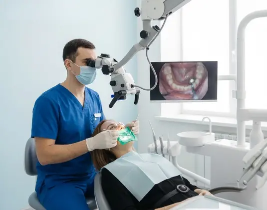
Microscopic dentistry: precision and reliability
High-tech dental treatment methods have become the norm today. Dental microscopy is the best proof of this. This dental treatment technique ensures exceptional precision and safety when performing complex procedures. The use of surgical optics with multiple magnification allows the dentist to see the smallest anatomical structures that cannot be seen with the naked eye or binoculars. Thanks to the use of a microscope, we can preserve and restore teeth that previously had to be removed.
Therapists at Club 32, a family dental clinic in the Holosiivskyi district of Kyiv, use microscope-assisted dental treatment for all complex clinical cases. We always make every effort to preserve your teeth and restore their functionality and aesthetics. Our specialists have many successful cases of using a surgical microscope to treat patients of all ages in their portfolio.
We practice an individual approach – we develop a unique plan for each patient, allowing us to perform treatment under a microscope, taking into account the specifics of the clinical situation and at an affordable cost. In addition, you can pay for treatment in installments and without overpaying.
We do everything to ensure that your treatment is effective, affordable, and comfortable.
Types of dental treatment under a microscope
The most popular areas in which dental microscopy is used include:
- endodontic treatment (treatment of the root canal or canals) under a microscope, aimed at cleaning, re-treatment, and filling root canals;
- removal of fragments of old fillings and broken instruments from root canals;
- detection and elimination of cracks, perforations, and canal obliterations;
- treatment of teeth with curved, narrow, or branched canals.
Each procedure is performed with precision, minimizing the risk of complications and increasing the long-term effectiveness of therapy.
Microscopic dental treatment procedure
The standard protocol includes:
Diagnostics using CT or high-definition radiography
This allows us to accurately determine the number, shape, and condition of the root canals, identify hidden inflammation, cracks, and anatomical abnormalities of the tooth, which is critical for successful treatment.
Cofferdam placement and antiseptic treatment
The rubber dam isolates the tooth from saliva and bacteria, creating a sterile field. Antiseptic treatment destroys pathogens on the surface and in the canal openings.
Mechanical and chemical treatment of the canals
The canals are widened with special instruments and rinsed with disinfectant solutions to completely remove infected tissue and pulp debris.
Use of ultrasound and irrigation with solution activation
Ultrasound helps dissolve biofilm and activate the antiseptic deep inside the canals, improving its penetrating ability and increasing sterility.
Sealing with biocompatible materials
The canals are filled with special composites (most often gutta-percha and cement), ensuring tight obturation without voids or overfilling.
Restoration of the anatomical shape of the tooth
After filling, the dentist restores the crown of the tooth with a composite or inlay, ensuring proper occlusion and protection from destruction.
Treatment of tooth canals with a microscope allows you to control every movement of the instrument and avoid complications such as perforation, missed canals, or ineffective obturation.
Advantages of treating teeth under a microscope
High accuracy of diagnosis and treatment
A microscope magnifies the image of the working field by 20-25 times. Therefore, microscopic dentistry dramatically increases the success of medical procedures even in the most complex and non-standard clinical situations. With the help of a microscope, the doctor can examine in detail the mouths of root canals, microcracks, areas of inflammation, and remnants of old fillings. Thanks to such detail, the doctor clearly understands where intervention is necessary and where healthy tissue needs to be preserved. This eliminates the "blind work" characteristic of traditional therapy.
The ability to preserve even severely damaged teeth
In cases of deep caries or advanced pulpitis, more than 50% of a tooth may be destroyed. When treated without a microscope, such teeth often have to be removed. But optical magnification allows even narrow and curved canals to be found, cleaned perfectly, and filled, preserving the supporting function of the tooth. This is especially important when treating molars and premolars involved in chewing.
Eliminating the risk of re-inflammation and tooth extraction
Untreated areas of infected tissue or missed canals are the main causes of unsuccessful endodontic treatment. A microscope allows you to miss no area. Monitoring each stage, from cleaning to obturation, reduces the risk of recurrent periodontitis, granulomas, cysts, and, as a result, tooth extraction.
Improved prognosis in complex cases
Complex cases include retreatment, instrument fragments, blocked canals, calcification, and anatomical abnormalities. Microscopic techniques allow these complications to be detected and eliminated without damaging surrounding tissues. This significantly increases the success of treatment even in "problematic" patients and minimizes the risk of complications.
Minimal interference with healthy tissue
The dentist can clearly see the boundary between affected and healthy tissue. This allows only damaged areas to be removed without affecting the structure necessary for the strength and restoration of the tooth. This approach increases the service life of the filling and reduces the need for prosthetics.
Economic feasibility
The price of root canal treatment under a microscope—the most common type of microscopic dental treatment—is naturally higher than similar treatment "blind." However, if you compare the cost of such treatment with the cost of tooth extraction, subsequent implantation, and prosthetics, the benefits become obvious.
Recommendations after root canal treatment under a microscope
Endodontic treatment under a microscope is a high-tech procedure, the long-term success of which depends not only on the professionalism of the dentist, but also on the responsible behavior of the patient.
Therefore, we strongly recommend that everyone who wants the results of treatment to last for many years follow these recommendations:
- avoid putting pressure on the treated tooth during the first 24 hours;
- use a soft brush and special rinses.
- do not delay permanent restoration.
- be sure to have a follow-up examination in 2–3 weeks.
If you are interested in dental treatment under a microscope in Kyiv, welcome to the Club 32 microscopic dentistry clinic. We employ highly qualified and experienced specialists who use the most advanced dental technologies.
We guarantee European quality, affordable prices, and friendly service.
Book a consultation now!
📞 (067) 382 32 32 ( 👉 Viber, 👉 WhatsUp, 👉 Telegram )
📍 Address: Kyiv, Stepana Rudnytskoho St 15, building 3
Instagram: instagram.com/club32.ua
Facebook: facebook.com/club32.ua